
INSIDE THE HEART

The human heart seen on the inside with front cut out. The four chambers of the heart are shown. Two top and two lower ones. To the right of the screen are the left sided structures including the mitral valve, papillary muscles, left ventricle and left atrium. To the left of the screen are the right sided structures including the right atrium, right ventricle, tricuspid valve and superior vena cava.
THIS IS THE HUMAN HEART

This image of the left lower portion of the heart or left ventricle show a normal mitral valve. The shape of the ventricle is still normal, different to the globular shape in some forms of mitral valve disease.
The human heart has four chambers: two on the left side and two on the right side. Unlike conventional wisdom, the heart sits mostly behind the sternal bone and slightly to the left. The reason we address the heart as the left and right is because each of these sides acts as pump. On the right side the heart receives the blood low in oxygen from the abdomen, legs, head, and arms pumps this blood to the lungs.
Once in the lungs, the blood exchanges the carbon dioxide for oxygen and then returns to the left side of the heart. It is here that the mitral valve plays an important role. The left side of the heart has two chambers, the left atrium (top) and the left ventricle (bottom). The blood that returned from the lungs goes into the left atrium first and then crosses the mitral valve to fill the left ventricle. When the left ventricle (real pump in the heart) contracts the pressure builds up and snaps the mitral valve shut preventing back leakage or regurgitation. The two leaflets of the valve close and touch each other (coapting) preventing the leakage. This simple one-way valve effect prevents the blood from backing up into the lungs, a phenomenon that would lead to high pressures in the lungs, and the enlargement of the left upper chamber (atrium), which in time leads to atrial fibrillation (arrhythmia). In the right side of the heart the ‘sister’ valve to the mitral valve is the tricuspid valve and functions in a similar fashion.
The Mitral Valve Apparatus

The mitral valve apparatus, as it is understood in medicine, consists of the valve leaflets or flaps (two: anterior and posterior), the chordae tendineae or simply chords, and the papillary muscles that act as anchoring points of the chordae to muscle of the heart.
The mitral valve as seen by a surgeon

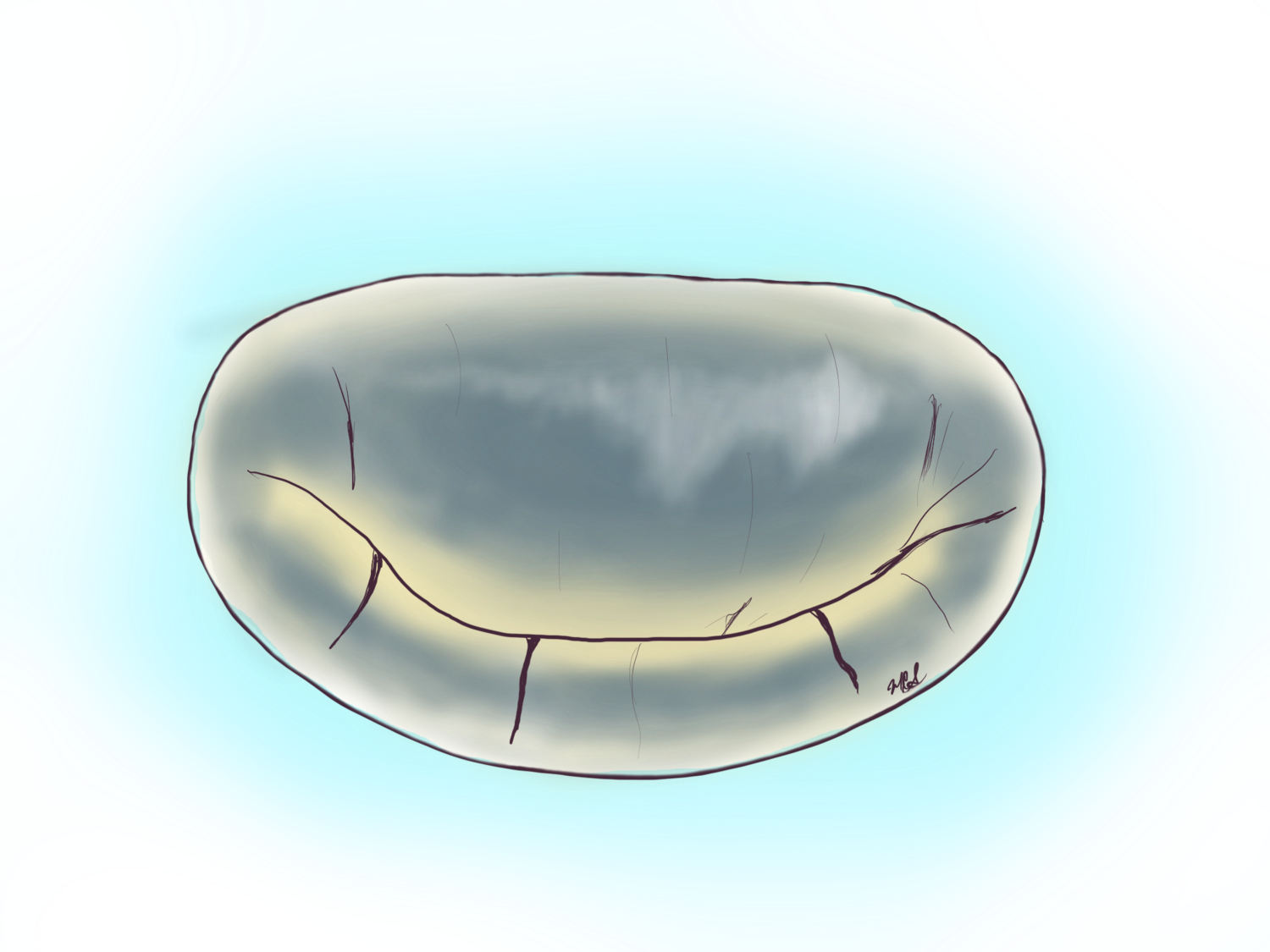
The mitral valve
The mitral valve as seen by surgeons as studied in the medical field. There are two leaflets of flaps to the valve, one anterior and one posterior. The valve is divided by imaginary lines into three anterior and three posterior segments. The frame surrounding the valve to which the leaflets attach is called the annulus. The leaflets are held anchored to the muscle of the heart through the papillary muscles.
The Role of the Mitral Valve
The mitral valve as a parachute

THe mitral valve as a parachute
The two flaps of the mitral valve called the anterior and posterior leaflets touch each other and seal the valve preventing blood from abnormally going upwards when the heart pumps. This is like the canopy of a parachute opening with air and being held down by the strings (chordae or chords in the mitral valve).
The mitral valve as a set of french doors

The mitral valve functions similar to a set of french doors. The image to the left shows in pink what is known as the annulus which would be the frame of the valve. The two leaflets or flaps of the valve would be the doors. These flaps should open only in one direction and the chords prevent them from flopping like saloon doors.

The mitral valve is a one way valve. When the valve closes the two flaps touch each other and prevent the blood from going upwards or leaking.
The Cardiac Cycle
The Cardiovascular System

The cardiovascular system has veins (above in purple) and arteries (above in red). Arteries carry the blood away from the heart to the different organs including brain, kidneys and heart itself. Veins return the blood from different organs back to the heart’s right atrium. The large artery leaving the heart is called the aorta. The largest veins arriving at the heart are the superior and inferior vena cava.
Both arteries and veins can be quickly accessed in the groin through a small incision.