
RHEUMATIC MITRAL VALVE DISEASE
the stenotic or tight mitral valve
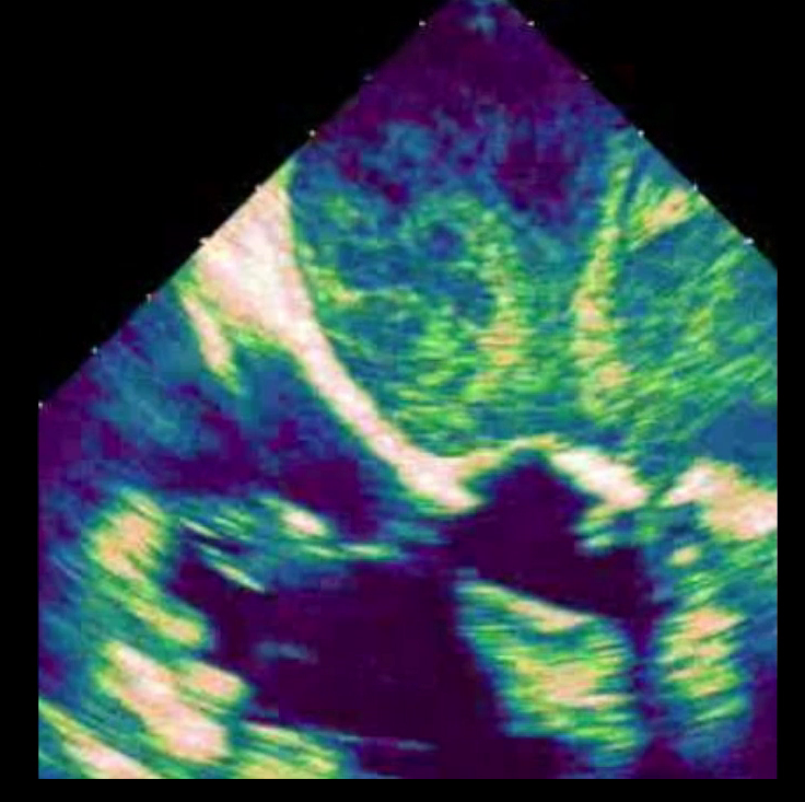
Mitral stenosis is usually from rheumatic heart disease. This is often accompanied by leakage. Two problems in one. This transesophageal echocardiogram shows the swirling pattern of the blood in the top part of the picture. This denotes the difficulty of the blood crossing the mitral valve and therefor higher pressure in the left atrium or top chamber.
Take Home Points:
Rheumatic mitral valve disease occurs after untreated rheumatic fever.
Rheumatic mitral valve disease can be mitral leakage (regurgitation) or mitral tightening (stenosis).
Management of rheumatic mitral regurgitation is often times mitral valve replacement and this can be performed minimally invasive.
In persons that suffered from rheumatic fever during childhood, typically secondary to strep throat infections that go unattended, a form of mitral valve disease may occur known as rheumatic valve disease. This is recognized as rheumatic mitral valve disease and is more frequently a situation where the valve does not open wide enough (mitral stenosis), but it can have regurgitation.
The disease process here causes a form of scarring of the valve tissue including the leaflets and chords, which in turn thicken and become stiff and poorly mobile. This form of mitral valve disease is more often treated with valve replacement. There are a few small size studies that supported repair of a rheumatic mitral valve; the evidence is still not sufficient to show the long-term durability of these repairs. In cases of rheumatic valve stenosis and regurgitation replacing the mitral valve is very acceptable option to correct the problem.
Rheumatic mitral valves resected at the time of replacement

This is an image of a rheumatic mitral valve showing clear signs of thickening and stiffness. There are also signs of heavy calcium deposit in to the left of the screen. This valve was leaking, but was also very tight.

Rheumatic valve disease is secondary to prior untreated infections by streptococcus, a bacteria. The valve in time thickens and many of the thin chords transform into what appear to be very thick pillars.
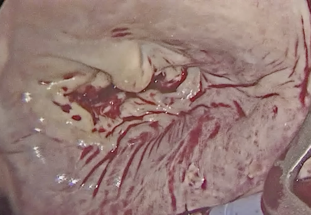
A picture of a rheumatic mitral valve at the time of minimally invasive mitral valve surgery. The valve is seen as a light yellow color in the center with some calcium deposits on it as well as thickened tissue.

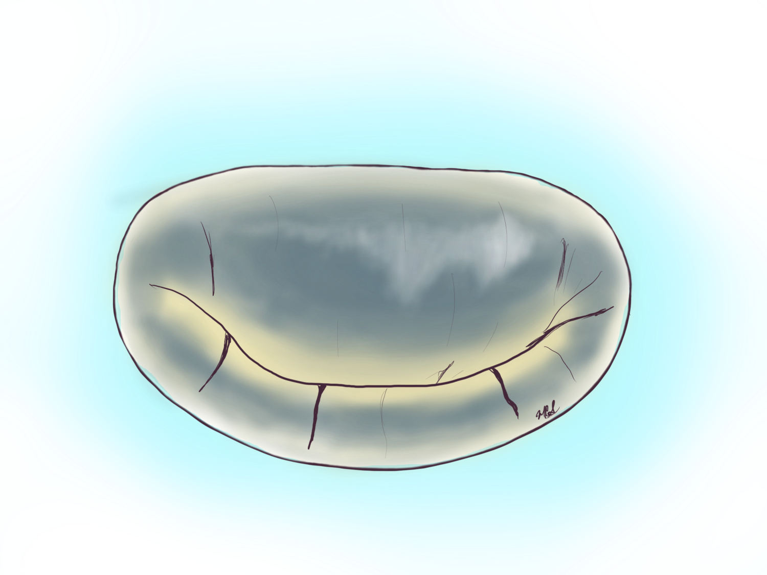
This is a trans esophageal echocardiogram of a mitral valve (white/grey structure around the blue color) with rheumatic disease. The opening of the valve is very small and leads to pressure backing up above it in the left atrium.