
The Leaking Mitral Valve or Mitral Regurgitation
The mitral valve may fail to close well and therefor leak. In general three levels of severity are found, mild, moderate, and severe. Patients with mitral valve regurgitation become symptomatic when the regurgitation is severe. The circumstances leading to severe mitral regurgitation vary from a slow build-up over years from mild to severe, to a fast and sudden event marked by shortness of breath.
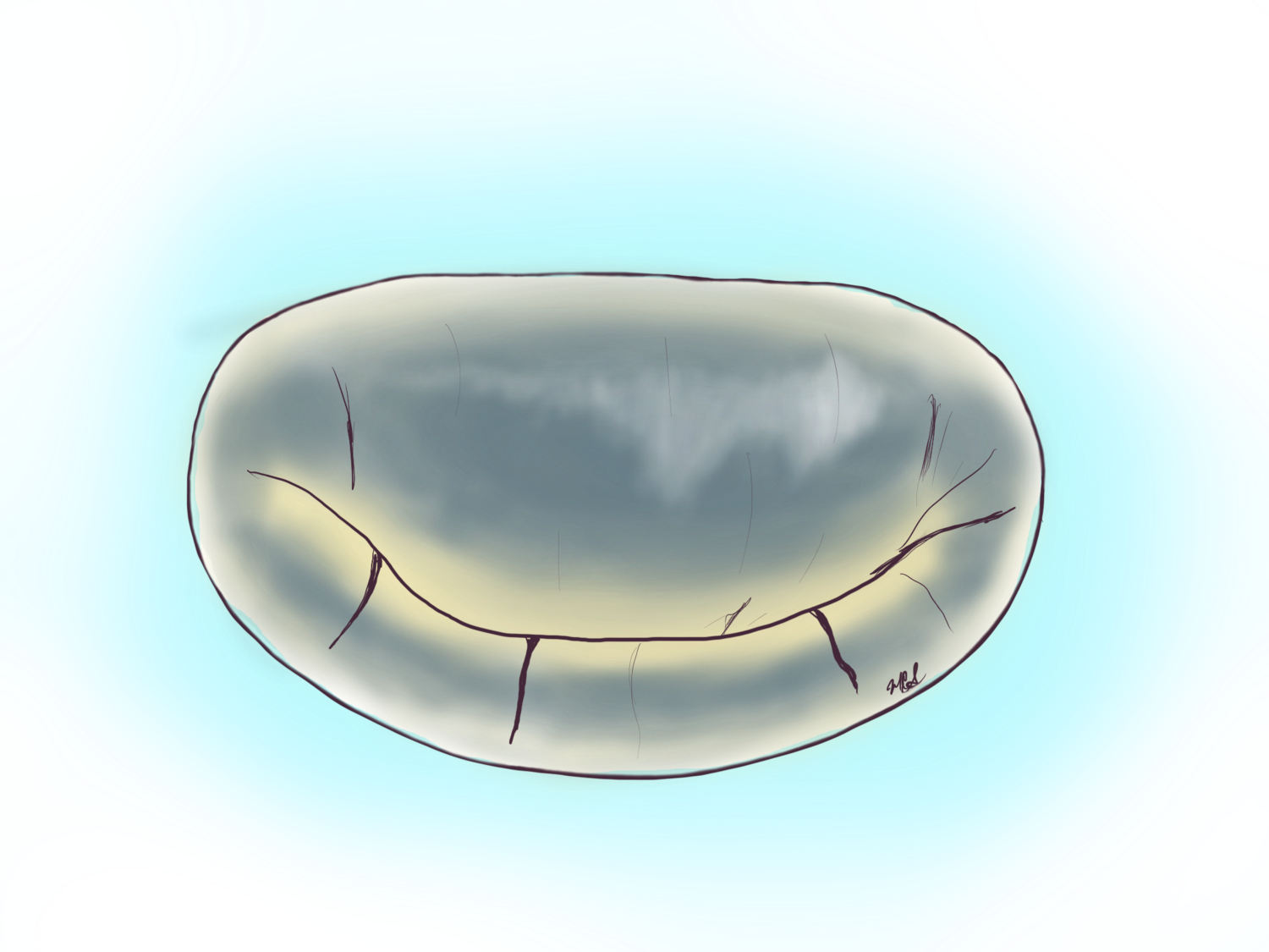
The Mitral Valve Apparatus

A cut-out of the mitral valve apparatus. To the lower right are the two papillary muscles that anchor the mitral chords to the leaflets or flaps of the valve. There are two flaps, one anterior and one posterior. Together all these structures are called the mitral valve apparatus. The papillary muscles are susceptible to ruptured and scarring from heart attacks, while the chords are susceptible to ruptured or stretching in some circumstances. The entire apparatus is susceptible to damage from bacterial infections and to the buildup of calcium.
Take Home Points:
Degenerative mitral valve disease is the most common form.
Valve repair surgery is the standard of care for degenerative mitral valve disease.
All forms of degenerative mitral valve disease can be repaired minimally invasive.
The mitral valve apparatus, as it is understood in medicine, consists of the valve leaflets (two: anterior and posterior), the chordae tendineae or simply chords and the papillary muscles that act as anchoring points of the chordae to muscle of the heart.
Examples of mitral leakage on echocardiogram
This transesophageal echocardiogram shows a restricted posterior leaflet with mitral regurgitation or leakage.
This transesophageal echocardiogram shows a damage to the posterior leaflet to the right of the screen leading to a large leakage directed to the left of the screen.
Why does the mitral valve leak?
Degenerative Mitral Valve Disease

Above is a spectrum of mitral valves within the category of degenerative mitral valve disease. The common denominator to this category is a damage to the valve tissue itself including the chords. Fibroelastic tissue disorder of the mitral valve may present as the simple rupture or stretching of the chords that hold the leaflets in place (prevent them from flopping backwards and causing leakage). In more advanced forms the material from which the leaflets or flaps are made stretch and become redundant, as the process progresses over the years the valve appears more redundant and more damaged. A severe form of this disease is characterized the most abundant amount of tissue with stretched-out chords or even ruptured chords, this form is known as Barlow’s disease.
The entire complex of problems leading this form of mitral regurgitation are often referred to as “Degenerative” mitral valve disease.
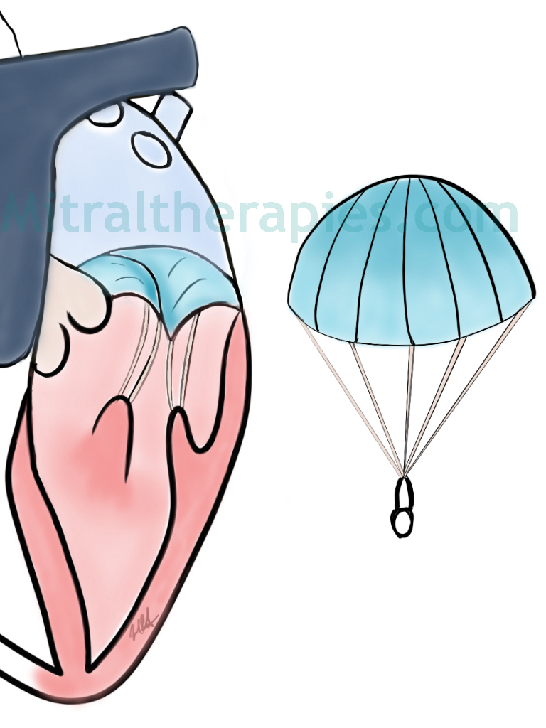
A simple analogy is that of a parachute with the canopy being the leaflets and the chords the strings that attach to two common points. In the same concept, if one of these chords is torn then the canopy will flail and wind will escape moving the parachute sideways. The same concept applies to the mitral valve. When a chord/s rupture the leaflet supported by these chords will flail and will allow blood to escape and leak. This phenomenon falls into the category of fibroelastic tissue disorder and is the most common form of mitral valve leakage. Together with this group there is a spectrum of disease severity ranging from the simple chordal ruptured to the more complex valves with redundant tissue, stretched-out chords the most severe form called Barlow’s disease. This form of disease that some call degenerative mitral valve disease is typically best treated with mitral valve repair as there is very good medical evidence that this is long-lasting.
Degenerative Damage to the Posterior Flap (Posterior Leaflet)

The most common form this form of degenerative disease of the mitral valve is when chords rupture or elongate in the posterior leaflet of the valve in the P2 segment. The second most common form is when both leaflets (anterior and posterior) show either elongated or ruptured chords.
Examples of posterior flap damage. Ruptured chords
posterior leaflet disease
This video shows a damaged mitral valve with ruptured chords to the posterior leaflet to the right of the screen. One can see the ruptured chords hanging from the edge of the posterior flap.
This trans esophageal echocardiogram shows a restricted posterior (or back) leaflet of the mitral valve causing regurgitation or leakage. The leakage is seen in yellow inside the triangle top right.
Degenerative Damage to the Anterior Flap (Anterior Leaflet)
The least common of the flails, for many years it was considered the most difficult of the mitral valve repairs to be performed. Today, with newer non-resectional techniques, and use of the techniques developed in Leipzig Germany this has become as easy to repair as the posterior leaflet pathology.
This video shows the pumping cycle of the heart with a damage to the mitral valve with ruptured chords (small thin strings) to the front or anterior leaflet of the mitral valve. The ruptured chords are shown hanging from the edge of the anterior flap to the left of the image.

This 3 dimensional image of the mitral valve at the time of a transesophageal echocardiogram clearly shows the ruptured chord on the top and middle of the picture (finger-like projection) and the elevation of the tissue at the top of the picture. The blood will leak under the raised flap, this is mitral regurgitation.
This two-dimensional echocardiographic picture shows the two flaps of the mitral valve: the anterior is to the left of the picture and the posterior is to the right. The anterior leaflet is flail or flopping in upwards above the level of the posterior flap (right).

This is the image of an anterior flap of the mitral valve that is flopping upwards. There is also a perforation or hole on the anterior leaflet. One can see the ruptured chords of the anterior flap (lower center).

Anterior leaflet of the mitral valve with ruptured chords. This is and example of degenerative mitral valve disease.
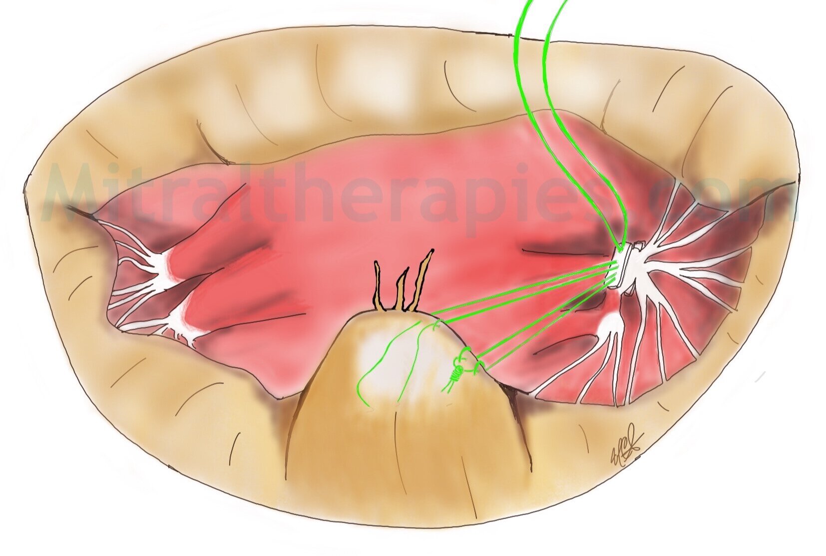
Degenerative Damage to Both Flaps (Bileaflet Damage)

When both leaflets of the mitral valve move upwards and leak this is known as bileaflet disease. If the chords are ruptured then both leaflets will flail, whereas if the chords are stretched this will lead to bileaflet prolapse. This form of disease has different degrees of damage and may require a combination of techniques to repair the valve including resectional and non-resectional techniques.
Illustration of damage to both flaps (bileaflet disease)
Echocardiogram of bileaflet damage
This two-dimensional transesophageal echocardiogram shows the prolapse or abnormal upwards rising of the mitral valve flaps (leaflets) that leads to the eventual separation of the two flaps leaving a gap between the two causing leakage for the mitral valve. This is considered "degenerative" mitral valve disease.
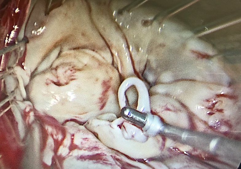
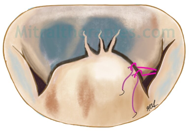
Degenerative Damage to the Corner Segments (Commissural Damage)
The corners of the mitral valve are known as commissures. The segments of the leaflets affected by ruptured or stretched chords can also be repaired to correct the leakage. This is not a common form of mitral valve damage.

Damage to the corner flap, in this case P3 third posterior segment. This damage was caused by ruptured chords which lead to leakage.

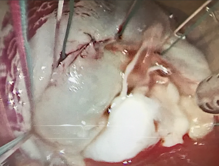
This picture taken at the time of mitral valve repair minimally invasive shows the medial or right sided commissure leaking from ruptured chords of the P3 segment. This valve was repaired.

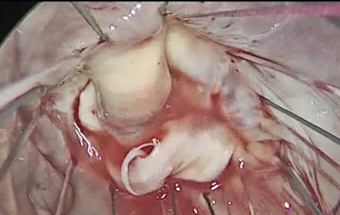
The same mitral valve from the prior pictures with a right sided commissure repaired through a minimally invasive approach.

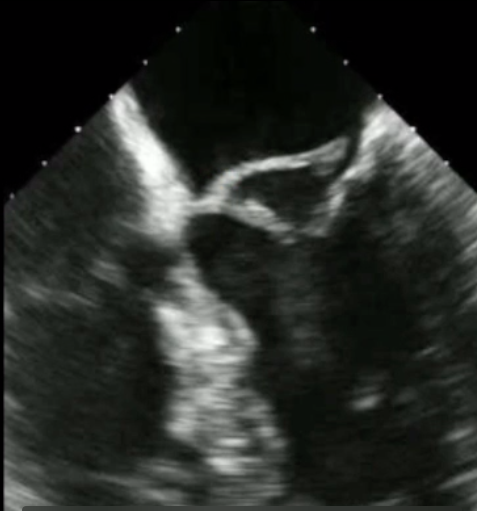
A tranesophageal echocardiogram showing the left ventricle on the right side with the corner flap riding higher than the anterior flap (see gap).

This same mitral valve on transesophageal echocardiogram showing the flow of blood from the left to the right top corner through the gap created at the corner segment.
Degenerative Barlow’s Disease of the Mitral Valve

A drawing of a mitral valve affected by Barlow’s disease.
Barlow’s disease of the mitral valve is a severe form of degenerative disease also known myxomatosis degeneration of the valve. In this form of disease the valve tissue degenerates and becomes excessive and redundant. The chords also stretch and may rupture too. Barlow’s disease does cluster in families, and although the exact genetic mechanism of inheritance is not know, it can be passed on from parents to offsprings.