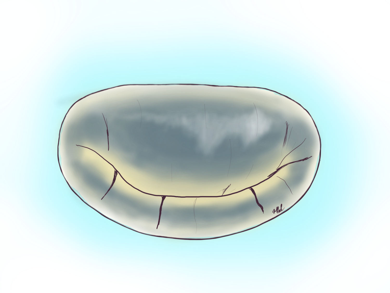
What is a new Murmur?
Take Home Points:
Murmurs are not normal in adults; a new murmur needs to be studied by a specialist.
A new mitral murmur warrants an echocardiogram and evaluation by a specialist.
A new murmur after an infectious illness raises suspicion for endocarditis.
Most new murmurs are diagnosed during routine visit to a primary care or internist. This finding warrants an echocardiogram, a non-invasive imaging study of the heart that allows us to see inside the heart cavities and assess the function of the valves within the heart. This study will be able to assess if a valve is not working properly as well as the severity of the problem. A murmur is a whooshing sound created by the passage of blood across an abnormal valve creating turbulence, the blood may be going in the natural direction of flow through a tight valve or against the natural direction of flow through a leaking valve.
Mitral valve regurgitation severity can be gauged by echocardiogram. There are three levels, mild, moderate, and severe regurgitation. The criteria for different levels of severity are well standardized and are based on objective criteria, not opinions.
Any person diagnosed with a new murmur should undergo an echocardiogram and be referred to a cardiologist for further evaluation. The low cost of echocardiogram (sonograms of the heart) and their availability almost anywhere in the United States make them a high yield, non-invasive way of assessing the problem and quantifying it.
Example of mitral leakage on echocardiogram
This Transesophagel echocardiogram shows a damage to the posterior leaflet to the right of the screen leading to a large leakage in yellow, directed to the left of the screen. The jet of leakage in yellow is the culprit for the murmur heard. In an echocardiogram this is called the regurgitant jet.
If a diagnosis of mitral regurgitation is made in the moderate range or higher, consideration for a transesophageal echocardiogram (TEE) is warranted. A TEE is an advanced form of echocardiography that is performed by introducing a probe through the mouth into the esophagus and stomach to look at the heart in higher definition. The images obtained from the TEE can give specific information as to the nature of the valve damage and better describe the leakage. Most hospitals that offer cardiovascular care perform TEE’s, but because it requires intravenous sedation medication, it requires someone to transport the person receiving the TEE.
Example of posterior leaflet flail or ruptured posterior chords
This video shows a damaged mitral valve with ruptured chords to the posterior leaflet. The blood leaking backwards into the left atrium or superior heart chamber causes turbulence at the mitral valve level which is perceived as a murmur with the stethoscope. The ruptured chords are shown hanging from the edge of the posterior flap to the right of the video.
A new murmur after severe infectious illness…
In some instances mitral valve disease is secondary to severe systemic or widespread infection that then affects the heart valves. This is called endocarditis. Often times the person affected by this may have had a severe illness with hospitalization and intravenous antibiotics given, but it can also present after an event perceived as trivial like an infected tooth or molar. When one presents with fevers, and a new murmur on examination, suspicion for endocarditis should be high. The workup is very similar to that of a new murmur including echocardiogram and likely transesophageal echocardiogram, but will also add blood samples for bacterial cultures, all in a more urgent fashion.
A new murmur after a heart attack…
Some persons that suffer a massive heart attack and do not seek medical attention may develop a complication from a heart attack. This complication is the damage or even complete ruptured of the papillary muscle. This leads to severe mitral regurgitation from the leaflets flapping upwards. This is a surgical emergency and requires a mitral valve replacement.

